Fig.7 Three-dimensional structure prediction and active site analysis of XmTPS protein A: Three-dimensional structure of XmTPS protein and its docking model with GPP B: Ramachandran plot analysis of the XmTPS protein model C: Prediction of active sites in XmTPS
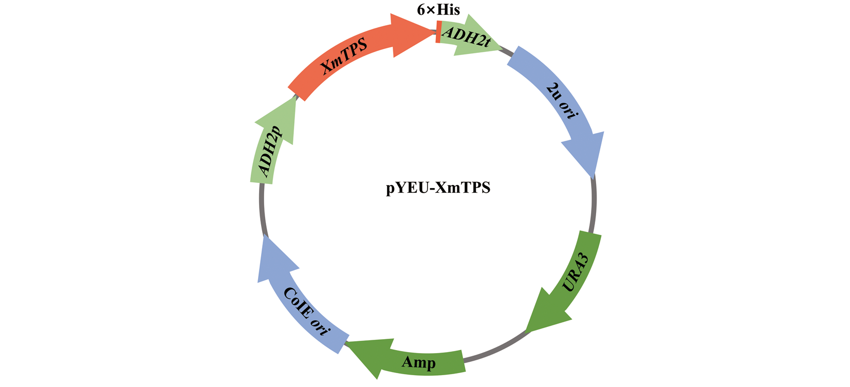
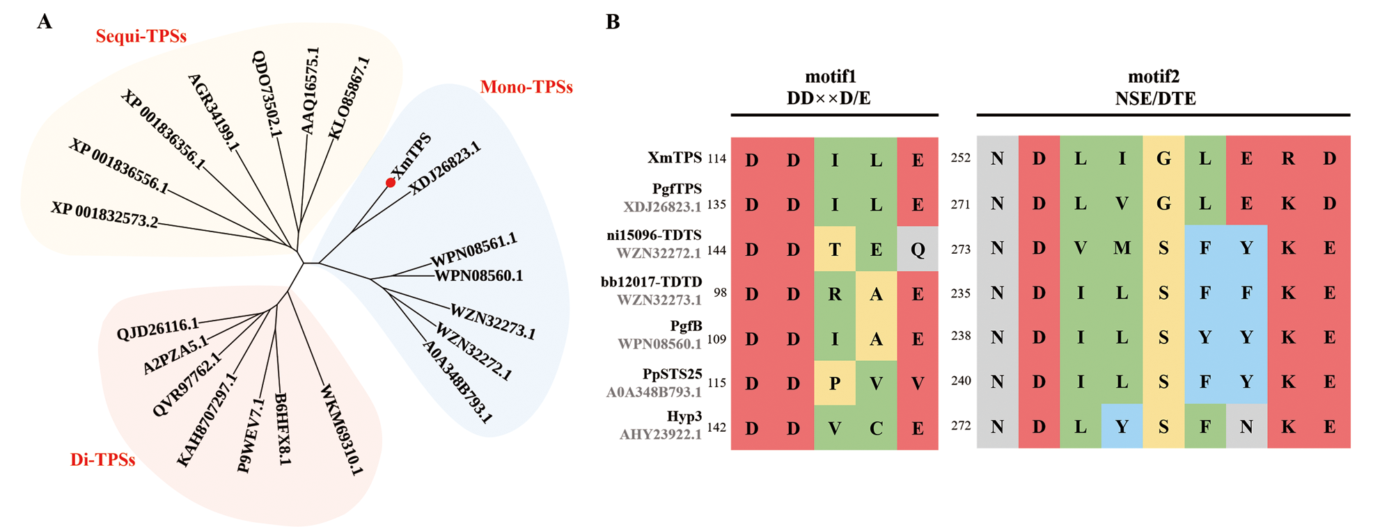
Other figure/table from this article